Focus Ion Beam
(Thin section FIB sample prep for Scanning Transmission Electron Microscopy)
What Is It?
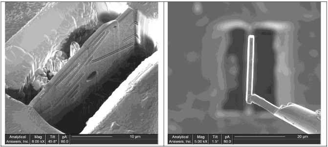
The Focused Ion Beam (FIB) technique is analogous to Scanning Electron Microscopy (SEM) in that it scans a focused probe beam, in this case ions rather than electrons, across the surface of interest.This beam can be used to generate high-resolution images of the sample or to mill into the sample to expose the internal structure.
Why Should I Use It?
This combination of high-resolution imaging to locate features of interest followed by precise site-specific milling provides an invaluable tool for sample preparation and analysis, providing rapid cross- sectional analysis of features that would be difficult or impossible to otherwise prepare.Because of this, the FIB instrument is often used as a sample preparation tool for more complete characterization using Electron Microscopy.
What Do I Get Out of It?
The combination of FIB and SEM/STEM is particularly powerful for characterizing the morphology and composition of layered structures and buried defects.
Applications Include:
- Process Development
- Protective coatings
- Inclusions
- Grain size distributions
- Layered structures
- X-sections of hard to polish
- materials
- Micromachining
- Failure Analysis
- Precise site-specific
- cross-sections
- Delamination
- ESD damage
- Sub-surface contamination/defects
- Quality Control
- Layer thicknesses
- Etch profiles
- Step coverage and conformality